
NeuXelec - SEEG analysis, simplified.
Research software

SEEG analysis, simplified.

From raw scans to precise electrode localization.
The complete SEEG workflow in one tool.

Zero scripting required
Data stays local
Free for research
100% Automated coregistration
2 clicks Per electrode reconstruction
30 min Per patient, imaging included
BIDS Compliant export standard

Everything a clinician needs
to work with implanted brains.

Project file

One .json per patient: image paths, electrodes, markers and camera positions in a single portable file.

Save & history

Clicking the logo (top-left) prompts to save and returns to the project selection menu. Clicking the (top-right) prompts to save and closes the application. Recent projects are listed at startup, sorted by last modified.

Two opening modes

Edit mode: full access. Visualization only: navigate and export, no modifications.

01

Import &
Coregistration

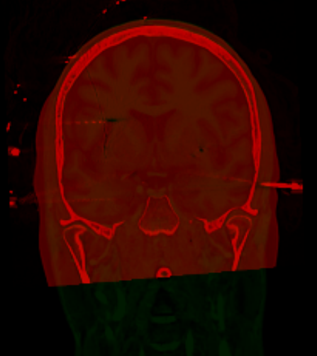
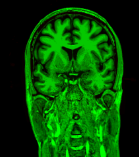
Load any combination of MRI 1, CT, MRI 2, PET and SPECT. NeuXelec aligns every modality to the MRI 1 reference automatically. No manual landmark picking, no command line.

  • NIfTI, DICOM & MGZ supported
  • ANTs-powered rigid registration
  • Automatic brain mask generation
  • One-click SISCOM computation
Explore in detail
T1 MRI CT PET
Aligned ✓
02

Electrode
Reconstruction

Click two contacts on the post-implant CT. NeuXelec reconstructs the full electrode trajectory and assigns precise coordinates to every contact. Minutes, not hours.

  • Simultaneous axial, coronal & sagittal views
  • Two-point automatic trajectory estimation
  • Per-electrode color & hemisphere labeling
  • Export to BIDS, TXT, CSV, TSV, JSON, Cartool ELS & iELVIS
Explore in detail
SEEG electrodes in 3D
03

Oblique &
Parcellation

Reslice any volume along the electrode shaft for a true contact-aligned view. Overlay CT, MRI, PET and SISCOM simultaneously. A live table maps each contact to its anatomical region.

  • On-the-fly oblique plane computation
  • Multi-modal opacity blending
  • FreeSurfer & custom atlas support
  • Automatic contact to brain region table
Explore in detail
Oblique + Parcellation Oblique + SISCOM Oblique + PET Oblique + CT
04

3D View
& Export

Visualize all implanted electrodes on the patient's cortical surface. Navigate coronal, axial and sagittal slices simultaneously, then normalize to MNI space for group-level analysis.

  • Hardware-accelerated 3D rendering (PyVista / VTK)
  • Coronal, axial & sagittal slice navigation
  • MNI152 normalization & coordinate export
  • Publication-ready screenshots & GIF export
Explore in detail
Pial surface rotation Electrodes on brain MNI atlas electrodes